Didymosphenia M. Schmidt in A. Schmidt et al.; 1899; 214
Key references
Krammer K., Lange-Bertalot H. 1986. Süßwasserflora Mitteleuropas. Bacillariophyceae. 1. Teil. Naviculaceae. Eds Ettl H., Gerloff J., Heynig H., Mollenhauer D., Gustav Fischer Verlag, Stuttgart. 876 pp
Metzeltin D., Lange-Bertalot H. 1995. Kritische Wertung der Taxa in Didymosphenia (Bacillariophceae). Nova Hedwigia. 60: 381-405.
Whitton B.A., Ellwood N.T.W., Kawecka B. 2009. Biology of the freshwater diatom Didymosphenia: a review. Hydrobiologia. 630: 1-37.
Metzeltin D., Lange-Bertalot H. 2014. The genus Didymosphenia M. Schmidt. A critical evaluation of established and description of 11 new taxa. Iconographia Diatomologica Vol. 25, Koeltz Scientific Books. 298 pp
Morphology
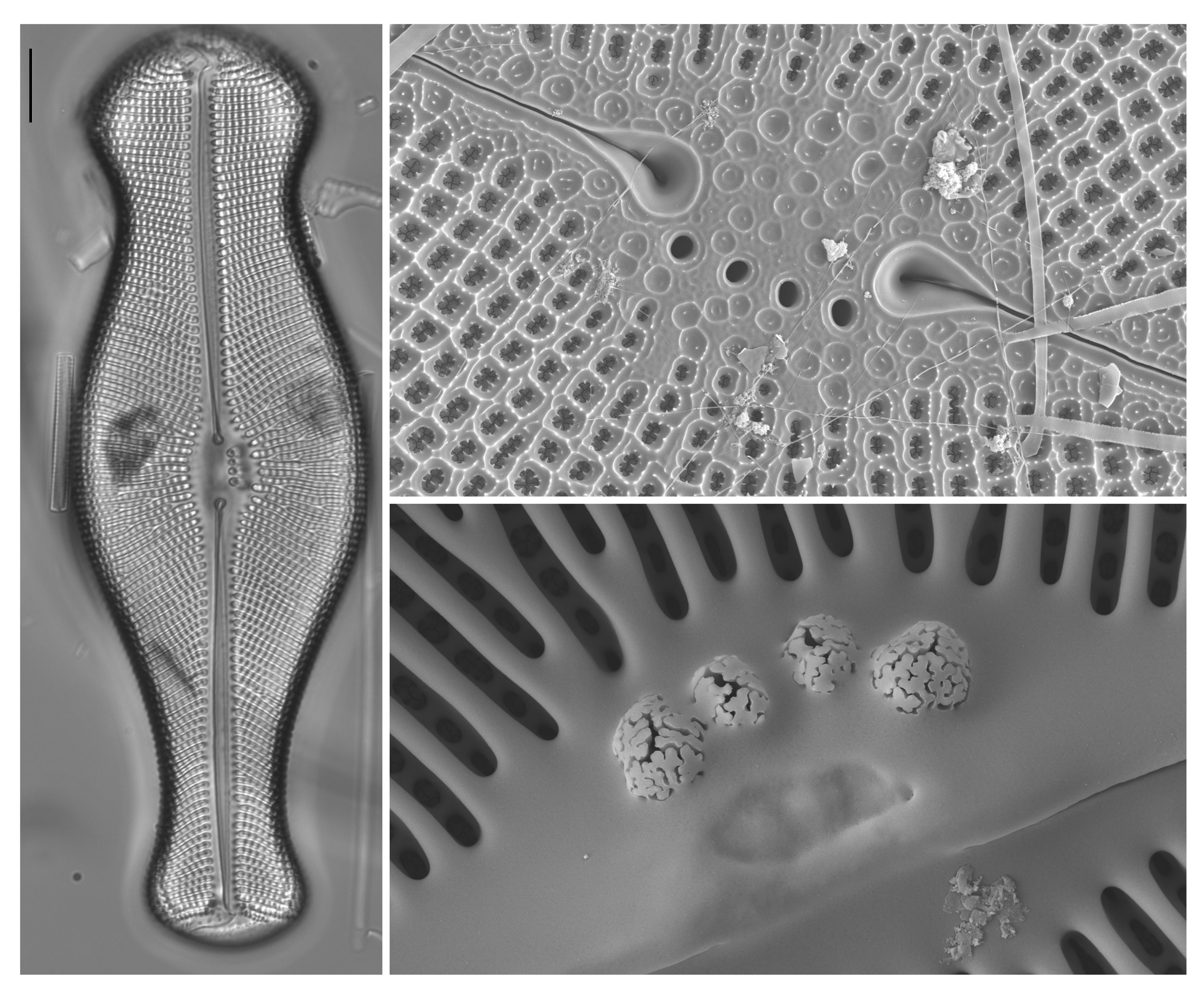
Frustules large, wedge-shaped in valve view, with a broad, bluntly rounded head pole, often a swollen centre and a rounded foot pole; wedge-shaped in girdle view; girdle deep (hence cells often lie in girdle view).
Frustules isovalvar, very slightly to noticeably asymmetrical with respect to the apical plane (with a tendency to be slightly ‘cymbelloid’).
Striae uniseriate, strongly radial with long and short striae alternating at the centre; areolae circular to quadrangular, very obvious in LM, occluded by complex flaps borne from the areola walls; a large area of many tiny pores is present at the base-pole, forming a pore field.
Axial area narrow, central area somewhat expanded, containing one to several isolated large pores (stigmata) on one side (the primary side).
Raphe central, straight or slightly curved; expanded externally at the centre to form raindrop-like endings; central internal endings hidden by silica overgrowth from the primary side; polar raphe endings bent towards the secondary side.
Girdle composed of several open bands. One complex chloroplast per cell, consisting of two X- shaped plates against the valves, connected by a substantial bridge on the secondary side of the cell (i.e. on the opposite side to the stigmata) containing a large pyrenoid.
Literature
References are given in chronological order.
Reference |
Citation |
|---|---|
| Schmidt A. 1899. Atlas der Diatomaceen-Kunde. Leipzig. O.R. Reisland. pl. 214; figs 1-12. | Morphology; Illustrations |
| Patrick R., Reimer C.W. 1966. The Diatoms of the United States. Exclusive of Alaska and Hawaii. Volume 1. Fragilariaceae, Eunotiaceae, Achnanthaceae, Naviculaceae. Monographs of the Academy of Natural Sciences of Philadelphia 13. 688 pp; 64 pls. | Morphology; Illustrations |
| Dawson, P. A. 1973. The morphology of the siliceous components of Didymosphaenia geminata (Lyngb.) M. Schm.. Br. phyco. J. 8: 65-78. | Morphology; Illustrations |
| Krammer K., Lange-Bertalot H. 1986. Süßwasserflora Mitteleuropas. Bacillariophyceae. 1. Teil. Naviculaceae. Eds Ettl H., Gerloff J., Heynig H., Mollenhauer D., Gustav Fischer Verlag, Stuttgart. 876 pp | Morphology; Illustrations; |
| Kociolek J. P., Stoermer E. F. 1988. A Preliminary Investigation of the Phylogenetic Relationships Among the Freshwater, Apical Pore Field-Bearing Cymbelloid and Gomphonemoid Diatoms (Bacillariophyceae). Journal of Phycology. 24: 377-385. | Morphology; Biology; Illustrations |
| Metzeltin D., Lange-Bertalot H. 1995. Kritische Wertung der Taxa in Didymosphenia (Bacillariophceae). Nova Hedwigia. 60: 381-405. | Morphology; Illustrations; |
| Blanco S., Ector L. 2009. Distribution, ecology and nuisance effects of the freshwater invasive diatom Didymosphenia geminata (Lyngbye) M. Schmidt: a literature review. Nova Hedwigia. 88(3-4): 347-422. | Ecology |
| Whitton B.A., Ellwood N.T.W., Kawecka B. 2009. Biology of the freshwater diatom Didymosphenia: a review. Hydrobiologia. 630: 1-37. | Morphology; Ecology; Taxonomy; Biology; Illustrations |
| Metzeltin D., Lange-Bertalot H. 2014. The genus Didymosphenia M. Schmidt. A critical evaluation of established and description of 11 new taxa. Iconographia Diatomologica Vol. 25, Koeltz Scientific Books. 298 pp | Morphology; Type Illustration; Taxonomy |
| Khan-Bureau D., Morales E.A., Ector L., Beauchene M.S., Lewis L.A. 2016. Characterization of a new species in the genus Didymosphenia and of Cymbella janischii (Bacillariophyta) from Connecticut, USA. European Journal of Phycology. 51(2): 203-216. | Morphology; Taxonomy; Illustrations; Biology |
This page should be cited as:
Mann D. G. Didymosphenia M. Schmidt in A. Schmidt et al.; 1899; 214. In: Jüttner I., Carter C., Cox E.J., Ector L., Jones V., Kelly M.G., Kennedy B., Mann D.G., Turner J. A., Van de Vijver B., Wetzel C.E., Williams D.M..
Freshwater Diatom Flora of Britain and Ireland. Amgueddfa Cymru - National Museum Wales. Available online at https://naturalhistory.museumwales.ac.uk/diatoms/browsespecies.php?-recid=3708. [Accessed:
].
Record last modified: 27/12/2020


