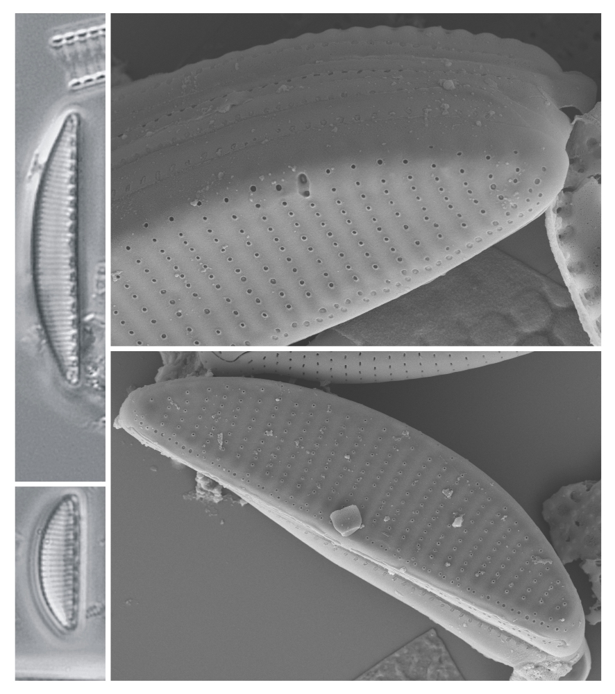
Cymbellonitzschia Hustedt in A. Schmidt at al.; 1924; pl.352
Key references
Krammer K., Lange-Bertalot H. 1988. Süßwasserflora Mitteleuropas. Bacillariophyceae. 2. Teil. Bacillariophyceae, Epithemiaceae, Surirellaceae. Eds. Ettl H., Gerloff J., Heynig H., Mollenhauer D., Gustav Fischer Verlag, Stuttgart. 596 pp
Cocquyt C., Jewson D.H. 1994. Cymbellonitzschia minima Hustedt (Bacillariophyceae), a light and electron microscope study. Diatom Research. 9(2): 239-247.
Morphology
Cells dorsiventral, with convex dorsal margin and straight ventral margin.
Frustules isopolar, isovalvar. Cells seen in valve or girdle view, rectangular in the latter. Raphe marginal, always lying on the ventral side in the most commonly encountered species (C. diluviana), which therefore exhibits ‘hantzschioid’ symmetry. In contrast, the type species (C. minima) exists as both hantzschioid and nitzschioid frustules (the latter having the raphe system on the dorsal in one valve and the ventral side in the other).
Striae for the most part uniseriate, with small, round areolae occluded by fine pore plates (hymenes) near their external apertures; the pore plates are more or less domed; in C. diluviana, the striae become biseriate and the areolae are smaller in the walls of the subraphe canal.
Axial area very narrow throughout and may bear a longitudinal ridge externally next to the raphe.
Raphe system fibulate, lying at the junction of the valve face and ventral mantle; the raphe itself is invisible in LM, its presence made apparent by the presence of fibulae, which are small square or rectangular structures; central raphe endings straight unexpanded; terminal fissures always bent towards the ventral side in species with consistent hantzschioid symmetry.
A few open bands per theca. Two simple plate-like plastids arranged ‘fore and aft’ (i.e. one towards each pole = on each side of the transapical plane), as in Nitzschia.
Cells solitary or forming short chains linked via their valve faces (but with no siliceous linking structures), attached via the ventral side.
Literature
References are given in chronological order.
Reference |
Citation |
|---|---|
| Schmidt A. 1924. Atlas der Diatomaceen-Kunde. Leipzig. O.R. Reisland Series. pl. 352; figs 12, 13. | Morphology; Description; Taxonomy; Illustrations |
| Krammer K., Lange-Bertalot H. 1988. Süßwasserflora Mitteleuropas. Bacillariophyceae. 2. Teil. Bacillariophyceae, Epithemiaceae, Surirellaceae. Eds. Ettl H., Gerloff J., Heynig H., Mollenhauer D., Gustav Fischer Verlag, Stuttgart. 596 pp | Morphology; Illustrations; Taxonomy; |
| Jewson D.H., Lowry, S. 1993. Cymbellonitzschia diluviana Hustedt (Bacillariophyceae): Habitat and auxosporulation. Hydrobiologia. 269/270: 87-96. | Morphology; Biology; Illustrations |
| Cocquyt C., Jewson D.H. 1994. Cymbellonitzschia minima Hustedt (Bacillariophyceae), a light and electron microscope study. Diatom Research. 9(2): 239-247. | Morphology; |
This page should be cited as:
Mann D. G. Cymbellonitzschia Hustedt in A. Schmidt at al.; 1924; pl.352. In: Jüttner I., Carter C., Cox E.J., Ector L., Jones V., Kelly M.G., Kennedy B., Mann D.G., Turner J. A., Van de Vijver B., Wetzel C.E., Williams D.M..
Freshwater Diatom Flora of Britain and Ireland. Amgueddfa Cymru - National Museum Wales. Available online at https://naturalhistory.museumwales.ac.uk/diatoms/browsespecies.php?-recid=4192. [Accessed:
].
Record last modified: 27/12/2020


