Amphipleura pellucida (Kützing) Kützing; 1844; 103
Key references
Stoermer E.F., Pankratz H.S. 1964. Fine structure of the diatom Amphipleura pellucida. I. Wall structure. American Journal of Botany. 51(9): 986-990.
Stoermer E.F., Pankratz H.S., Bowen C.C. 1965. Fine structure of the diatom Amphipleura pellucida. II. Cytoplasmic fine structure and frustule formation. American Journal of Botany. 52(10): 1067-1078.
Morphology
Shape
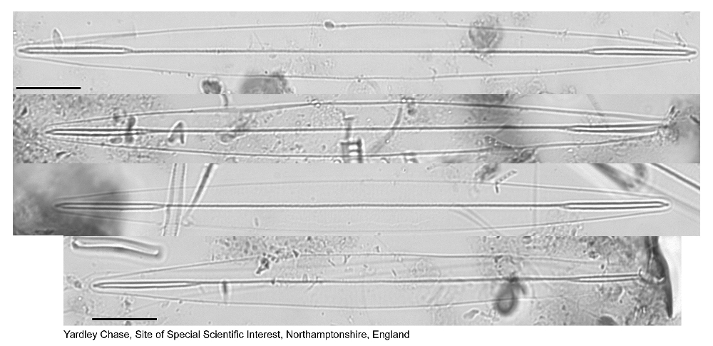
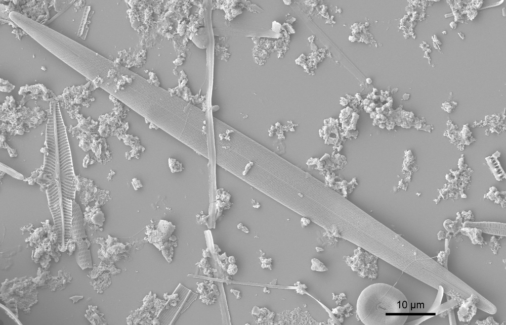
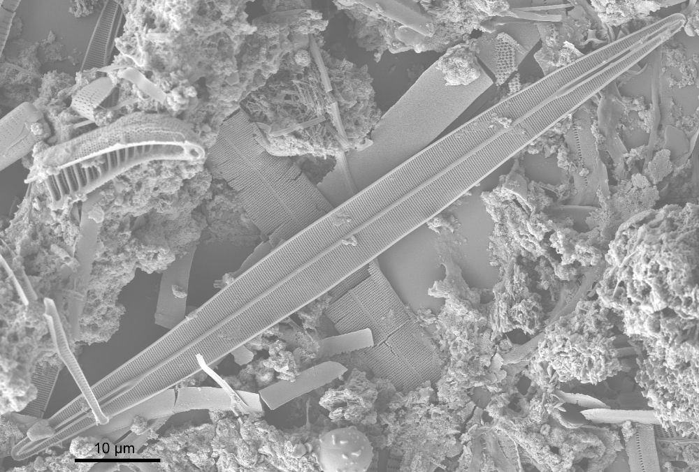
Linear-lanceolate valves with cuneate ends.
Symmetry
Isopolar, isovalvar, bilaterally symmetrical.
Striae
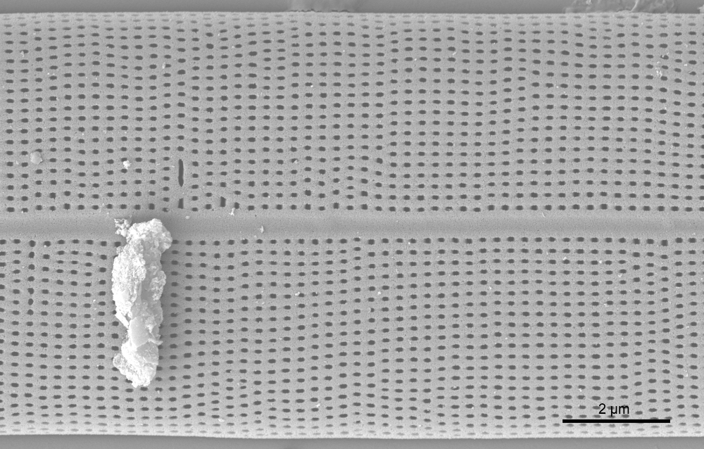
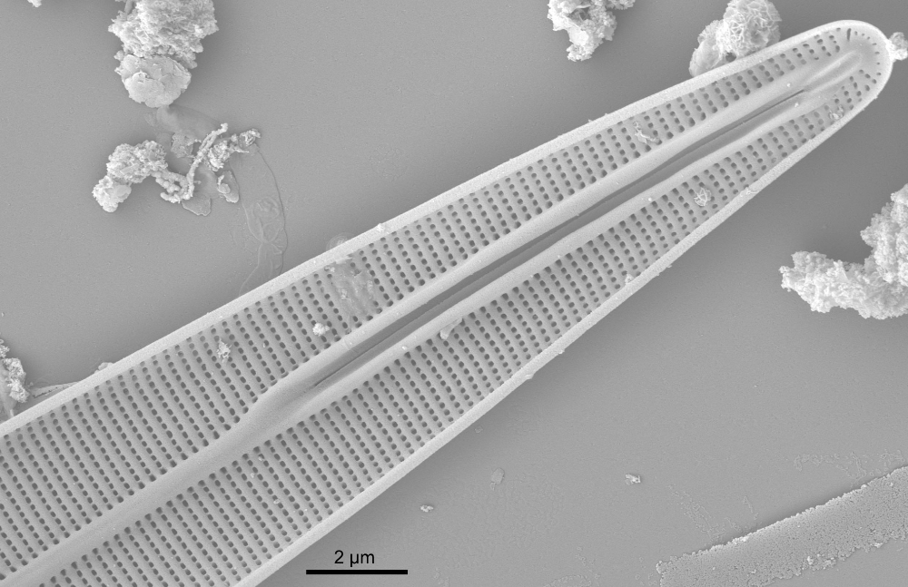
Very fine, not or hardly discernable in LM, uniseriate, perpendicular to raphe and parallel, composed of small areolae.
Raphe
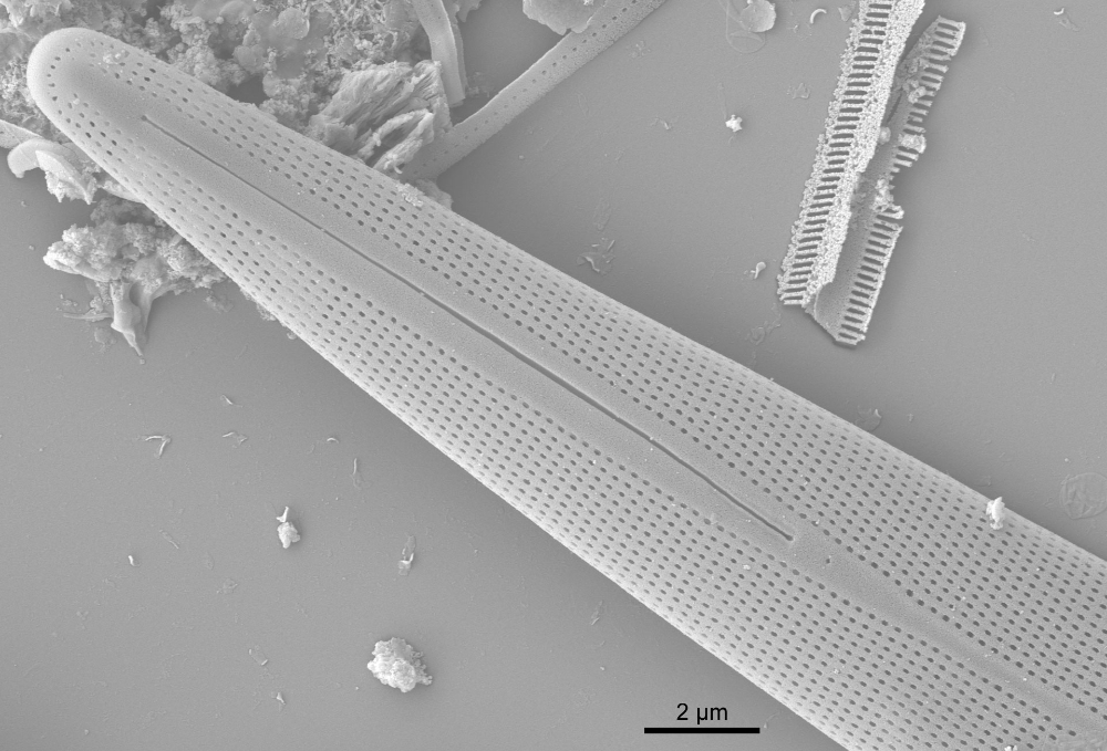
Two short raphe slits towards valve ends, separated by a long, narrow axial sternum.
SEM morphology
Internally the sternum projects as distinct rib that splits into two branches which enclose the raphe slits. Close to apex, the two ribs are fused with the helictoglossa to form the ‘porte-crayon’.
Literature
References are given in chronological order.
Reference |
Citation |
|---|---|
| Kützing F.T. 1844. Die Kieselschaligen Bacillarien oder Diatomeen. Fr. Fritsch, Nordhausen. 152 pp; 30 pls | Morphology; Description; Illustrations |
| Stoermer E.F., Pankratz H.S. 1964. Fine structure of the diatom Amphipleura pellucida. I. Wall structure. American Journal of Botany. 51(9): 986-990. | Morphology; Illustrations; |
| Stoermer E.F., Pankratz H.S., Bowen C.C. 1965. Fine structure of the diatom Amphipleura pellucida. II. Cytoplasmic fine structure and frustule formation. American Journal of Botany. 52(10): 1067-1078. | Illustrations; Biology; |
| Round F.E., Crawford R.M., Mann D.G. 1990. The Diatoms. Biology & Morphology of the Genera. Cambridge University Press, Cambridge. 747 pp | Morphology; Illustrations |
| Krizmanić J., Ržaničanin A. , Subakov-Simić G., Cvijan M. 2008. Amphipleura pellucida (Kütz.) Kütz. - an emended diagnosis concerning valve length. Diatom Research. 23(1): 243-248. | Morphology; Illustrations |
| Höbel P., Sterrenburg F.A.S. 2011. UV photomicrography of diatoms. Diatom Research. 26(1): 13-19. | Technology; Morphology |
| Sclafani M., Juffmann T., Knobloch C., Arndt M. 2013. Quantum coherent propagation of complex molecules through the frustule of the alga Amphipleura pellucida. New Journal of Physics. 15: 083004. | Technology; Illustrations |
| Lange-Bertalot H., Hofmann G., Werum M., Cantonati M. 2017. Freshwater Benthic Diatoms of Central Europe: Over 800 Common Species Used in Ecological Assessment. Cantonati M., Kelly M.G., Lange-Bertalot H. (eds.), Koeltz Botanical Books, Schmitten-Oberreifenberg, Germany. 942 pp | Morphology; Illustrations; Ecology |
This page should be cited as:
Kelly M., Jüttner I., Mann D. G., Jones V., Williams D. M. Amphipleura pellucida (Kützing) Kützing; 1844; 103. In: Jüttner I., Carter C., Cox E.J., Ector L., Jones V., Kelly M.G., Kennedy B., Mann D.G., Turner J. A., Van de Vijver B., Wetzel C.E., Williams D.M..
Freshwater Diatom Flora of Britain and Ireland. Amgueddfa Cymru - National Museum Wales. Available online at https://naturalhistory.museumwales.ac.uk/diatoms/browsespecies.php?-recid=4539. [Accessed:
].
Record last modified: 27/12/2020


