An Introduction to Shell Structures
Experience with students has shown us that the biggest factor preventing correctly identifying bivalves is a lack of familiarity with shell structures and terminology. There is no short-cut to gaining this knowledge although we try to present a much information visually.
General Features
The bivalve shell typically consists of two calcareous, convex valves that are hinged dorsally and free ventrally. The hinge margin is typically united by a non-calcified ligament and a set of articulating hinge teeth. The valves lie on the left and right sides of the animal. The shell grows comarginally around the post‑larval shell, which remains as a beak somewhere along the dorsal margin. The area surrounding the beak is known as the umbo although some regard these terms as interchangeable. Each valve can be regarded as having; dorsal, ventral, anterior and posterior margins. The beaks effectively divide the dorsal margin into anterodorsal and posterodorsal parts. If the beaks are situated medially the valves are equilateral but if the beaks lie infront of or behind the midline then inequilateral. The beaks may face each other across the dorsal margin, i.e. orthogyrate but more commonly they point in the anterior, prosogyrate or posterior opisthogyrate directions. In a few bivalves, they may actually be coiled.

Internal features of a bivalve shell

Regions of a bivalve shell
Many bivalves have areas demarcated by angles or ridges that radiate from the umbos. The most common of these is a posterior angle or posterior ridge running over the umbos to the posterior ventral margin. If the ridge is elevated it is known as the posterior carina. The area behind this angle is known as the posterior area or posterior slope. In some groups, a radial depression or groove, the posterior sulcus, may accompany the posterior angle. Less frequently and to a lesser degree there may be a similar anterior angle demarcating the anterior area.In between these angles lies the median area. In a few forms, especially those with a large byssus, the median area may be depressed to form a median sulcus.
Two important features are found on the dorsal margin. Anterior to the beaks there may be a defined, often heart shaped area, the lunule. Posterior to the beaks there may be a similar structure which is usually elongate in outline and depressed, the escutcheon.

Dorsal features of a bivalve shell
In many bivalves the valves do not meet at all points around the margins. These gapes vary in position and most commonly occur posteriorly where it reflects the presence of large siphons. An anterior or anteroventral gape is usually for the extension of the foot and is referred to as the pedal gape. Many bivalves use a byssus extruded from the foot to attach themselves to their substrate. If the byssus is large there is often a ventral or byssal gape.
Scallops and scallop-like forms such as Lima have shells that possess ear like extensions, auricles, of the dorsal margin. In Pteria the auricles may be greatly extended into wings. In scallops the valves remain on the right and left sides although the animal lives in the opposite plane lying on one valve. The byssus is accompanied by a notch in the right valve, and often with a series of small teeth, the ctenolium, along the ventral margin of the notch.

In strongly inequilateral shells the posterior dorsal edge can be further divided. In mytilids the ligament is greatly extended along the dorsal margin and this portion termed the ligament margin can he differentiated from the remaining dorsal edge by a change in angle or curvature.
In cemented bivalves orientation varies between families but in the oysters, the only group represented in our fauna, they are always attached by the left valve.
In many species the valves are not the same size or may not be the same shape. This, inequivalve, condition may be extreme as in Corbula or subtle as in tellins.
Top of pageHinge Ligament
The ligament joins the valves along the hinge margin, it is elastic, mostly brown or black in colour and serves to open the shell when the adductor muscles relax. It is an important diagnostic character but is a complex structure and often difficult to interpret.
PositionThe ligament nearly always lies between or posterior to the beaks. It may be easily visible when the valves are closed “external” partly visible “sunken” or not visible “internal”. Some ligaments have both external and internal components.
OrientationThe ligament may lie symmetrically between the beaks “amphidetic” or more often behind the beaks “opisthodetic” and very occasionally in front of the beaks “prosodetic”
Structural types
There are four main types.
 |
 |
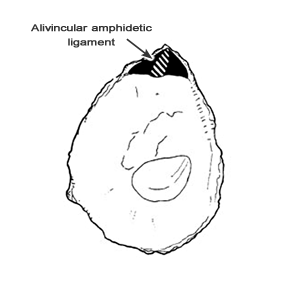
| “Alivincular” is a flattened, usually triangular, structure lying between the beaks on a flat cardinal area and composed of a central fibrous layer bounded by lamellar layers on either side. External alivincular ligaments are typical of Limopsis and Ostrea.
If the cardinal area becomes deeply cleft the ligament becomes internal as in the Pectinoidea. |
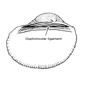
“Duplivincular” is where there are multiple bands of fibrous and lamellar ligament inserted on the cardinal area, usually forming v-shaped chevrons as in the Arcidae and Glycymeridae but may be aligned vertically as in the Noetiidae. |
 |
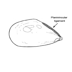 |
| “Parivincular” is an arched structure lying behind the beaks. Typical of Veneridae and Tellinidae | “Planivincular” an elongated weakly arched ligament extending posteriorly as a narrow band. Typical of the Mytiloidea. |
Calcareous structures associated with the ligament
“Resilifer” a depression in which the ligament is attached. Most often used for the attachment pit of internal ligaments. Can be seen as a shallow triangular pit in the cardinal areas of Limopsis and Lima.
“Chondrophore” the term applied to an internal resilifer that is expanded into a spoon shaped or platform-like structure that projects beyond the hinge plate. Typical of Mya and Scrobicularia.
“Lithodesma” a calcareous structure that supports an internal ligament. Often lost in disarticulated valves, typical of Lyonsia and Cuspidaria.

“Nymph” a dorsally projecting ridge that supports an external parivincular ligament. Typical of venerids and tellinids
“umbonal crack” a natural fracture of the shell close to the umbo caused by stress related to ligament growth, only in Cochlodesma.
Hinge Teeth
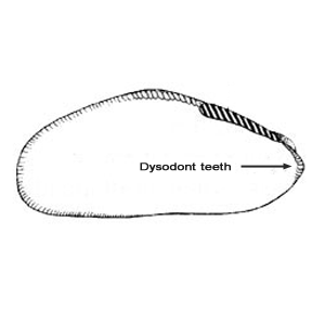
The articulating dorsal margins of each valve are usually thickened “hinge plate” and bear an assortment of teeth or ridges, collectively termed “hinge teeth”. If none are developed the hinge is said to be “edentulous”. In those forms with teeth, their form, number and disposition are frequently of systematic significance. There are four basic forms of hinge.
 |
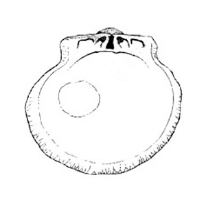 |
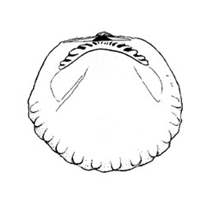
| “Taxodont” The teeth are numerous usually increasing in number with growth, all taking the form of simple projections and situated in a row along the dorsal margins. Typical of protobranchs, Arcidae and Limopsidae. | “Isodont” This hinge has only a few teeth placed symmetrically either side of the ligament. The teeth are ridge like or rectangular and are called crura. They may interlock so well that the valves cannot be separated without breaking the teeth. Here only in the Anomioidea but best seen in the Spondylidae. |
 |
 |
| “Dysodont” This describes the condition where there are no true teeth, only a few small, ill-defined denticles situated either side of the ligament. They resemble the marginal denticles of many bivalves and are found most frequently in the Mytiloidea. | “Heterodont” This is the hinge form found in the majority of bivalves. The teeth are few in number and differentiated in two forms. Cardinal teeth, which number from 1‑3 in each valve, radiate from the beaks. Lateral teeth, which number from 1‑2 either side of the cardinal set, are situated at some distance from the beaks and are subparallel to the shell margin. The combinations of cardinal and lateral teeth are great and attempts have been made to classify and formulate these. These are, however, not straight forward and are not included here. Confusion can also arise through fusion of some teeth, or through erosion. The latter is important in some lucines and tellins where some teeth may be represented only by denticles or weak ridges. In fragile shells, hinge teeth are easily fractured and care should be taken to note if there are any fracture lines present. |
Muscle Scars
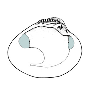
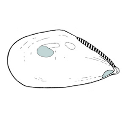
Most bivalves exhibit scars on the interior of the valves that result from the attachment of muscles. These reflect the gross anatomy of the animal and are important in classification.
“Adductor muscles” typically two, situated medially close to the anterior and posterior margins serve to close the valves.

“Anterior adductor and posterior adductor scars” impressed scars in the shell typically circular to oval, rarely elongate or crescentric in outline.
 |
 |
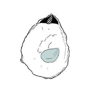 |
| “Dimyarian” where both adductors are present. the condition is termed, and “Homomyarian” where adductors are of equal size. | “Heteromyarian” where adductors are unequal in size. In groups that become sessile and attached by a byssus the anterior portion of the shell is often reduced, resulting in the diminution of the anterior adductor. | “Monomyarian” where only a single adductor is present. |
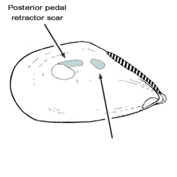
“Pedal muscles” Most bivalves possess a foot that can be used for burrowing or crawling and from which the byssus is extruded. The foot is attached to the shell by a number of “pedal protractor and retractor muscles”. In burrowing homomyarian forms he pedal muscle scars are small but in byssally attached heteromyarian forms the “posterior pedal retractor scar” is large and significant. This scar is situated close to the posterior adductor scar. In taxa such as Arca, Pteria and Pinna it is also employed as a “byssus retractor” but in mytilids and Anomia a separate byssus retractor is present. The relative size and positioning of pedal and bysul muscle scars can be very important in separating closely related species of mytilids, giant dams and anomids.
One of the most important features of the bivalve shell is the “pallial musculature” that is associated with the attachment of the mantle to the shell. The mantle tissue, which secretes the shell, is attached by a series of muscles close to the margins of the shell.

“Pallial line” a linear scar known runningclose to the edge of the shell, occasionally may take the form of an interrupted series or may be added to by accessory scars.
“Entire” where the pallial line follows the margins without any indentations.
“Pallial sinus” a posterior indentation of the pallial line reflecting the development of the siphons and may be shallow or extend right up to the anterior adductor scar.
The description of he pallial sinus is important. It can be said to have dorsal, anterior and ventral sections, if the ventral section is fused with the pallial line it is “confluent” if not then free. The dorsal section may be ascending (directed towards umbo) or descending (towards ventral margin). The anterior section may be rounded (sinus will be oval), straight (sinus will be rectangular), or the dorsal section may meet the ventral section directly (sinus will be acute). For the ventral section other than the point of confluence, it is also important to note the angle at which it meets the pallial line.
“Cruciform muscles” a pair of muscles that act in the control of the siphon and which often leave two small circular scars close and ventral to the end of the pallial line. Present only in tellins but often obscure.
Top of pageValve Orientation
Right or left?
To distinguish right from left valves requires discerning the anterior and posterior margins. Once established if the shell is held with the hinge margin uppermost and the posterior and anterior in the same plane as the obrever then the right and left valves will be in the right and left hands of the observer.
In shells with a external or sunken parivincular (eg. venerids, tellinids, lucinids) or planivincular (eg. mytilids) ligaments this always lies posterior of the beaks.
In shells with a pallial sinus this is always posterior, this is especially useful in taxa with internal ligaments.
In taxa with a byssal notch (eg scallops and Pteria) this is always anterior.
In rostrate shells the narrowed and extended end is usually posterior.
In shells witht an alivincular or duplivincular amphidetic ligament (eg. limopsids and glycymerids) orientation is best done from a knowledge of the internal anatomy. In limopsids the posterior adductor is larger than the anterior, in glycymerids the converse applies.
In general the mouth, labial palps and tip of the foot are anterior whereas the anus, rectum and siphons are posterior.
Orientation in nuculids and many galeommatids is difficult, many being slightly extended anteriorly, reference to the anatomy is the most reliable.
Dimensions
“Length” Maximum dimension along the longitudinal axis, anterior to posterior
“Height” Maximum dimension along the vertical axis, dorsal to ventral
“Tumidity” Maximum inflation of articulated valves, often termed breadth but this often confused with length.
“Thickness” Actual thickness of the shell of single valve, a measure of robustness or fragility.
Outline
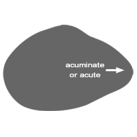
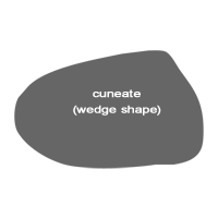
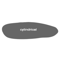
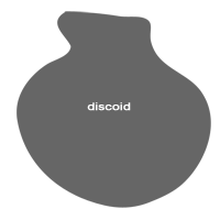
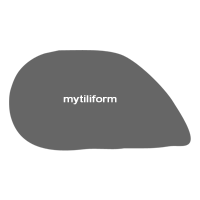
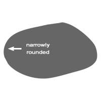
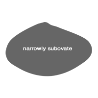
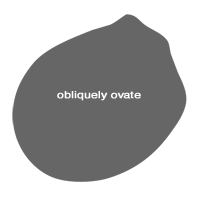
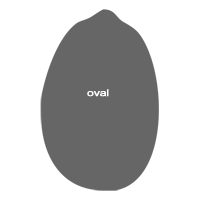
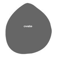
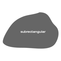
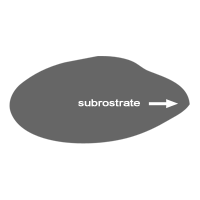
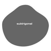
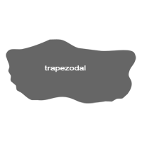
The outline of the valves is variable and often subtle. Unlike leaf shape in botany the many forms have no absolute definition.
 |
 |
 |
 |
 |
 |
 |
 |
 |
 |
 |
 |
 |
 |
 |
 |
 |
 |
 |
 |
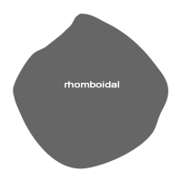 |
 |
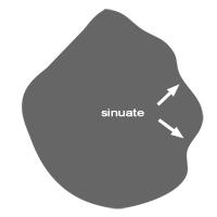 |
 |
 |
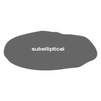 |
 |
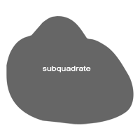 |
 |
 |
 |
 |
 |
 |
Sculpture
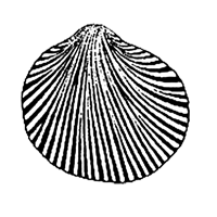
Shell sculpture is a widely used character for identification of species but requires careful observation and description. The prominence of any sculpture is variable such that terms of “weak” or “strong” are only part of a continuum. Numerical definitions can also be variable with numbers of a feature often increasing with growth. The following definitions give a guide to the terminology used here but may not agree exactly with others.
Sculpture consists of two broad elements:
 |
 |
| "Radial” any linear pattern that originates at the beaks and radiates towards the margins. | “Co-marginal” or “Concentric” any linear patterns that follows the margins. |
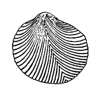
Other less common patterns that do not conform to the above are:
 |
 |
| “Oblique” or “acentric” where the linear feature is angled across the shell and does not radiate from the beaks. | “Scissulate” dense oblique striations often abruptly angled to concentric striations, seen in many tellins |
 |
 |
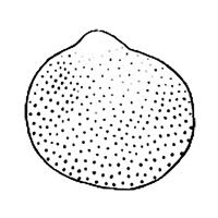 |
| “Divergent” where radial elements diverge from their normal orientation and where secondary elemens appear. | “Divaricate” where divergent radial element are angled in opposite directions, the angulations in a line medially or post-medially. | “Non linear” where the shell is wholly or partly textured, if raised then granular or pustulose, if sunken then pitted. |
Radial Patterns
The strength of raised radial sculpture varies from the faint to the strong thorough a series termed Lines – Threads – Riblets - Ribs
 Lines Lines |
 Threads Threads |
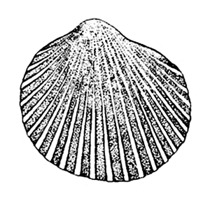 Riblets Riblets |
 Ribs Ribs |
Incised scupture is usually very fine “Striations” or fine “Grooves”
Secondary ribbing
In many shells the ribs are simple and the number of ribs remains thte same throughout growth. In many others the first ribs “primary ribs” can divide or new ribs “secondary ribs” can appear between the primaries, this process can be repeated to give rise to “tertiary elements”.
Cross sections and spacing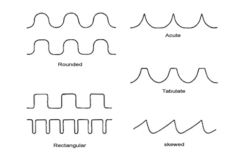
Ribs have a variety of cross sections
Rounded - Rectangular - Acute - Tabulate – Skewed
And the spacing can be equal to, wider than or narrower than the ribs.
Concentric patternsThe strength of raised concentric sculpture varies from the faint to the strong thorough a series termed Lines – Lirations – Ridges – Undulations.
The projection also varies from thin low lamellae to strong foliations
Cross sections and spacing

Cross sections vary from broad low rounded ridges to down-curved or up-curved lamellae to vertical foliations
Combined patternsIn many species the sculpture is composed of both radial and co-marginal elements, sometimes the radial is dominant with the co-marginal forming a variety of structures on the riblets or ribs. In others, the co-marginal is dominant and in others, they are equally developed.
“Cancellate” where the radial and co-marginal elements intersect to give a surface of regular roughly rectangular blocks
“Decussate” where the radial and co-marginal elements intersect to give a surface of angular blocks, generally finer than cancellate.
“Imbricate” where the radial elements are crossed by an interrupted series of co-marginal scales.
“Reticulate” where the surface has a fine network of intersecting raised threads.
“Cross bars” narrow ridges set at right angles on radial ribs
“Tubercles” rounded raised structures set on radial ribs
“Scales” lamellar structures arising from radial ribs, may be erect or flush like tiles.
“Spines” erect pointed structures arising from ribs.
